Tuesday, March 25, 2008
Aeolosomatidae and Potamodrilidae
Monday, November 12, 2007
The Polychaeta...
Length of polychaete species range between 3 m to less than 1 mm; the smallest wpecies are those of the genus Diurodrilus (about 300 um). There are several families which are exclusively meiofaunal: Dinophilidae, Diurodrilidae, Nerillidae, Protodrilidae, Protodriloidae, Saccocirridae, Polygordiidae, Parergodrilidae, Psammodrilidae, nearly all Psionidae and many Syllidae are meiobenthic.
Tuesday, October 02, 2007
The Oligochaeta...
Meiobenthic oligochaetes occur in a great variety of aquatic habitats. In freshwater, they are largely confined to the coarse sands and gravels of lakes and river beds or associated with the vegetation. In any given area, the sublittoral oligochaetes can generally be divided into two assemblages, one characteristic of muddy bottoms and one, often richer in species, in coarse sands and gravel.
Saturday, September 15, 2007
Copepoda Harpacticoida: General Structure (Part 4)
Males are nearly always smaller than females but are also distinguished by the structure of the antennules, genital somite and the P5 (which is smaller and less ornate than the females)
Monday, July 09, 2007
The Kinorhyncha
The number and arrangement of neck and trunk plates is addition to the number and arrangement of spines and adhesive tubes constitute the basis for further classification of the 100 species whose descriptions are based on adult specimens: nearly 60 additional species have been described from juvenile stages which likely will be unrecognizable with an adult.
The Kinorhyncha are divided into 2 orders, the Cyclorhagida and the Homalorhagida. There are three habits in which Kinorhynchs are found. Most are found in the upper 0-3 cm organically enriched mud to muddy sand estuaries, or the sea, from intertidal to abyssal depths. Some are found in the association with algae or other invertebrates. The third habitat is the interstices of medium to coarse marine sand or gravel: either intertidal sand of high energy beaches ort subtidal medium to coarse sand, “shell-gravel”, and “shell-rash”, “Amphioxus-sand” or even “Halimeda-sand”.
Wednesday, July 04, 2007
Copepoda Harpacticoida: General Structure (Part 3)
The mandibles also have a 2-segmented protopod and are biramous. The endopod is tipically 1-segmented rarely 2-segmented) and the exopod has a maximum of 4 segments. Reduction in obth rami, but usually the exopod, can result in a uniramous, 1-segmented mandibular palp.
The maxillules have a praecoxa with several spines and setae around its distal margin and usually with two setae on the anterior surface (absent in some reduced forms).
The maxillae are uniramous (the exopod absent). The endopod is at most 4- segmented.
The maxillipeds vary considerably throughout the order.
Tuesday, June 19, 2007
About Nematoda (Part 4)
The cuticle is the most external part of the body wall in nematodes. It is an elastic, proteinaceous, layered secretion of the epidermis. Four main layers can be distinguished: epicuticula, exocuticula, mesocuticula and endocuticula. The last three are also called cortical, median and basal layers.
The cuticle covers the entire body surface of the nematode and penetrates inside through various openings such as mouth and pharynx (stomodeum), anus and rectum or cloaca (proctodeum), vulva and vagina, excretory pore, external sensilla and glands. The body cuticle is semi-permeable and metabolically active. Liquids can pass thorugh it and pore-canals have been demonstrated in a number of species. At the same time the cuticle forms a barrier that protects the nematode against biotic and abiotic factors from its environment.
The ornamentation of the cuticle is one of the most important caracteristics in the identification of a nematode and usually is easy to be seen in light microscopy. There are many variations of the ornamentation however the three most common cuticle types in marine nematodes are:
Smooth:

Striated:

Pointed (with dots):
Saturday, June 02, 2007
Copepoda Harpacticoida: General Structure (Part 2)
The cephalosome bears six (seven if it is a cephalotorax) pairs of appendages? Antennules, antennae, mandibles, maxillules, maxillae and maxillipeds. A rostrum usually projects forward between the antennules from at the anterior margin of the cephalic shield.
Saturday, May 19, 2007
The Priapulida
Prior to 1968, the phylum Priapulida was considered to be a cold-water, macrobenthic taxon, represented in the meiofauna only by larval stages. The 7 species and 4 genera were al placed in a single family. Beginning with the discovery of Tubiluchus carallicola from the shallow, warm waters of Curaçao, the number of pripaulid taxa has steadily increased to 15 species, 7 genera, and 3 families. Most (7 species) or all (8 species) of these new taxa are meiofaunal. However, they are not merely miniatures of their larger macrofaunal relatives. They exhibit many new morphological and ecological characters demonstrating that the basic priapulid body plan is much more plastic than it was once thought to be.
The Priapulida are vermiform marine invertebrate, their body is unsegmented, cylindrical, and generally has three body regions: an introvert, abdomen, and tail or caudal appendage. Fourteen of the 15 species of extant priapulids are included in the three families: Chaetostephanidae, Priapulidae and Tubiluchidae. Meiofaunal priapulids include all species of the families Chaetostephanidae and Tubiluchidae and are generally associated with abundant and diverse meiofaunal assemblages. This is in marked contrast to be macrofaunal priapulids which appear to most common in sediments with a poor fauna. Though all meiofaunal priapulids, with the possible exception of the tube-dwelling Maccabeus appear to be active burrowers like the larger macrofaunal species.
Saturday, March 03, 2007
About Nematoda (Part 3)
A common nematode presents 2 body openings (excretory pore and anus/cloaca) for males and juveniles and 3 (excretory pore, vulva and anus) for females.
All the openings are in the ventral region helping in the determination of left and right sides of the body. This information is very important for the correct positioning of diverse structures (i.e. teeth).
On the slides, when correctly mounted, the animals are layed down on one of laterals (left or right) permitting the visualization of the body openings.
The body openings determine the body regions:
Pharyngeal region: from the mounth to the excretory pore
Median region: between the excretory pore and the anus (or cloaca)
Tail region: from anus (or cloaca) to the tail end.
The tail format is important to distinguish some genera and its length is one of the measures used to diferenciate and describe species!!!
In this figure can be seen the openings and the regions of a common nematode body:
Tuesday, February 27, 2007
The Loricifera...
The loriciferans are the most recent phylum described. The first known representative of this exclusively marine meiobenthic taxon was discovered in 1974 in medium sand at 400m depth off the coast of North Carolina. Since the description of the phylum nine species representing three genera and two families have been described, but over 23 other species are known currently. Adults loriciferans are bilaterally symmetrical and between 115-383 um long. The Phylum Loricifera consists of a single order, the Nanoloricida Kristensen, 1983, and two described families: the Nanolaricidae, consisting of two genera each with three species and the Pliciloricidae, with the genera Pliciloricus and Rugiloricus.
Tuesday, February 20, 2007
Phylum Rotifera
As the name Rotifera suggests, one of their most diagnostic features is the corona or “Wheel”-like complex of cilia in the head region which functions is locomotion and feeding. They are commonly vermiform metazoans, sometimes with elaborate cuticular spines or ornamentation, but rarely exceeding 2mm in length and more commonly less than 0.5 mm long. An estimated 2,000 species of rotifers have been described and divided into three classes, five orders up to 22 families and 123 genera and subgenera. Ten of the families have meiobenthic representatives. Although the majority are known from freshwater, rotifers are also found in brackish water and in the sea.
The Rotifera are composed of three classes. The class Pararotatoria is represented by a single genus, Seison, an exclusively marine taxon epizoic on the gills of the crustacean Nebalia. The remaining two classes include the Digononta and the Eurotatoria: the latter class contains most of the known species of the phylum.
Monday, January 08, 2007
Gastrotricha
Members of this phylum are small, strap or tenpin-shaped, acoelomate worms. Most adults are less than 1 mm in length, some species are less than 100 um, and others exceed 3 mm. The body is flattened ventrally and arched dorsally. It is divided into two regions, the head and trunk which are often externally indistinct. The head bears the mouth, a tubular nematode-like pharynx, and the brain. The trunk contains a straight tubular gut and the reproductive organs, and bears the inconspicuous ventral anus. All gatrotrichs have ventral locomotory cilia and, with them, glide smoothly over the substratum. When disturbed they adhere with adhesive papillae, which are often numerous.
Gastrotrichs can be distinguished from other vermiform ciliated micrometazoans by a combination of features. They glide smoothly over the substratum and do not typically writhe from side to side as is typical, for example, of flatworms. The gastrotrich mouth is anterior or, terminal, or slightly subterminal unlike that of most turbellarians and gnathostomulids in which it is ventral. The gastrotrich pharynx resembles that of nematodes, with which they are not easily confused, and is unlike those of turbelarians, annelids, and gnathostomulids. The larger gastrotrichs, which include most macrodasyidans and a few chaetonotidans, are simultaneous hermaphrodites. Small species, which include most chaetonotidans, reproduce by parthenogenesis although sometimes vestigial testes may be present. As far as is known, hermaphroditic gastrotrichs copulate as reciprocally cross-fertilize.
Tuesday, January 02, 2007
Nemertina
The phylum Nemertina is comprised of non-segmented worms whose anatomy includes: the dagnostic rhynchocoel housing a protrusible proboscis, a closed blood-vascular system lined by endothelium, a regionated gut with anal pore, a fully ciliated and glandular epidermis, a nervous system with four cerebral ganglia and a pair of lateral nerve cords, and serial sacculate gonads. There are about 900 described species of nemertines. They are primarily marine and found in all major marine habitats, but they also are known from freshwater and terrestrial habitats. The size of adult worms ranges from 1 mm to many meters in length and 0.15 mm to a few centimeters in width. The phylum consists of two classes, the Anopla and Enopla, the names reflecting respectively the absence or presence of specialized proboscis armature the stylets.
Interstitial nemertines generally are less than 0.5 mm in diameter and may be up to 50 mm in length, but many are less than 0.3 mm in diameter and 10 mm in length. About thirty species of interstitial nemertines have been described. Of the three orders, the hoplonemertines predominate in taxonomic diversity with 23 species in five genera.
Saturday, December 23, 2006
Gnathostomulida
Since their description and recognition as a separate phylum, Gnathostomulida have been encountered worldwide as na often abundant constituent od marine meiofauna. Most members of this phylum are of meiofaunal size and live in the interstitial spaces between sediment particles. At present, there are 18 genera with over 80 known species, but many more can be expected. The phylum is unique in having a body epithelium in which each cell carries only one cilium, and a bilaterally symmetrical muscular pharynx, equipped with cuticular mouth parts. Gnathostomulida are worm-shaped, cylindrical or with a slight dorsal-ventral flattening, and range from 300 to 3000um in length and 50 to 100 um in diameter; however, the most frequently observed genera have a body width of 50 to 70 um. The body is divided into a head which is commonly expanded and well delimited, a long trunk which constitutes 90% of the body, and a relatively short tail.
All gnathostomulids are hermaphrodites. The phylum is divided into two orders, the elongated Filospermoidea (with filiform sperm: without paired sensory organs, and without a bursa, vagina, or penis stylet), and the usually more compact Bursovaginoidea (with sperm not filiform; and usually with paired siensory organs and a bursa). Representatives of the phylum have been encountered in all the world’s oceans, from the supratidal brackish groundwater to the intertidal, and the subtidal to 800m. As a rule they occur in fine to coarse sand with a high admixture of organic detritus as typically found in sheltered bays, intracoastal waterways, between coral reefs, or wherever slightly reducing conditions may exist.

Wednesday, December 20, 2006
Copepoda Harpacticoida - General Structure (Part 1)
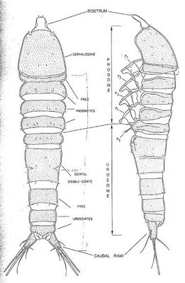
In the sub-class copepoda, there are two basic plans of body organization or tagmosis, gymnoplean and podoplean, differentiated by the position of the major body articulation. In the gymnoplean plan, this is behind the fifth pedigerous somite whereas in the podoplean plan (Harpacticoida) it is between the fourth and fifth pedigerous somites. The major articulation divides the body into an anterior prosome and a posterior urosome. The prosome is further into two sub-regions. Anteriorly is the cephalosome bearing the five head appendages and the maxillipeds and posterior to this are three free prosomites bearing the second to fourth swimming legs. The urosome comprises an anterior somite bearing the fifth pair of legs and five other somites (referred to as abdomen). In the males, all the usoromites are separated but, in the females, the second and third urosomites are usually fused to form the genital double-somite. The last urosomite or anal somite, in which the median anus opens either terminally or dorsally, bears two posterior caudal rami or furcal rami.

Tuesday, December 12, 2006
Turbellaria
Because of their postion among the simplest of metazoans, the “Turbellaria” have always received considerable attention concerning thier comparative morphology and systematics and the inter-relationship of these with their phylogeny or the evolution. However, may still represented a useful facies of a predominantly freeliving flatworm with a ciliated epidermis distinguishable from the similar Gnathostomulida by, among other things, the lack of a pair of jaws and base plate in the mouth.
Turbelarians are ubiquitous forms in freshwater and marine habitats; they are very associate with other organisms to various degrees (some are parasistic). About 2/3 are small (1 – 2 mm long or less), but osme, the marine polyclads and terrestrial triclads in particular, can grow to many centimeters in length. There are three main turbellarian life cycles types: univoltine, bivoltine, and polyvoltine. Among which have either continuous or discontinuous sexual maturity throughout the year. Univoltine and bivoltine life cycles are predominantly found in all other taxa. Turbellarians are very common on and in various benthic substrates (soft bottom, sandy bottom, calcareous bottom, gravel, mussel associations, vegetation) both intertidally and subtidally. Some turbellarians live in ground waters and caves in the stygobios.
Friday, December 08, 2006
About Nematoda (Part 2)
Most of the free-living Nematoda has a vermiform body shape, turning to fine on the extremities, the head and the tail.

However some nematodes present different shapes.
Fusiform: typical for the sub-order Desmoscolecina

"S" format: typical for the family Draconematidae.
Another curiosity about this family is the larger region anteriorly which looks like a head but in fact is the pharyngeal region.

“з” format (epsilon): typical for the family Epsilonematidae

Different body shapes indicate special patterns of locomotion as can be seen in the figure:

Concerning the size, free-living nematodes varies from 82μm (Greffiella minutum) up to some centimeters (some spp. of Enoplida).
Sunday, December 03, 2006
Ciliophora and Cnidaria
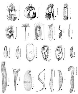
The ciliates, some 8,000 described species, comprise the phylum Ciliophora, of the Kingdom Protista and are cosmopolitan in distribution. These heterotrophic protests are characterized by two very distinctive features, nuclear dualism – diplod micronuclei and polyploidy macronuclei – and a unique infraciliature or kinetidal system, consisting of single or paired kinetosomones with closely associated microtubules, microfibrils, are various specialized organelles. Most individuals are ciliated over at least a portion of the body during some stage in the life cycle. Possession of pellicular alveoli is another feature characteristic of most ciliates. Feeding structures in these protests vary considerably between groups, with some forms lacking any while others possess very elaborate oral apparatuses. Replication is usually via binary fission of a homothetogenic nature, essentially transverse or perkinetal (across the kineties or rows of cilia). Ciliates have been described from a wide range of habitats, both marine and freshwater as well as terrestrial: and they may occur as free-swimming or sessile or sedentary forms, while still others are ecto- or endosymbionts of a diversity of hosts.
Ciliates play an important role in the food web, serving as prey for many metazoa and as predators (grazers) of/on bacteria. Ciliates act as nutrient recyclers for their bacterial prey.
The phylum Cnidária is characterized by only two layers of their body wall, the ectoderm and the endoderm, separetd by a generally thin acellular jelly layer, the mesogloea. This layer become extensive in the mecusae. All cnidarians are equipped with cnidocists (nematocysts) of different types, which mainly have the function to catch prey but may be used for defense and in in fixing vagile specimens to substrates. The species may be solitary or colonial. Many of them secrete an outer skeleton, which may be the chitinous periderm in Scyphozoa, Cubozoa and hydrozoa or the calcareous exoskeleton in Anthozoa and some hydrozoa.
All four classes of the Cnidaria are represented in the meiofauna: two species in the Scyphozoan, four in the Cubozoa, one in the Anthozoa, and possibly 28 species in the 15 genera in the Hydrozoa. Actually, most cnidarians live in coarse sand and shell gravel, more or less vigorously moving around in the interstitial space. However, some species occur in muddy sediments or in detritus rich surface layers.


Friday, December 01, 2006
Sarcomastigophora
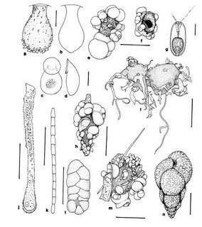
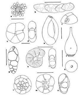
Foraminífera occur in all marine environments and a few species penetrate into brackish or fresh water. They are normally most common in fine-grained sediments. In general, they are less abundant in coarse sands without some silt and clay than in muds, presumably mecause food is scarcer. Many sediment-dwelling foraminifera live on or near the sediment surface where the nutrients are concentrated and the pore-water well aerated. Some species occur infaunally, usually in the upper few centimeters of sediment but sometimes deeper, while others mey be both infaunal and epifaunal.